US Scientists Develop Groundbreaking 3D Printing Technique for Biomechanical Research
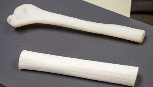
Scientists at UT Southwestern Medical Centre have discovered a new 3D printing process that enables the manufacture of realistic human femur models, marking a significant leap in biomechanical research. The Journal of Orthopaedic Research published the study, which is the first to validate 3D-printed femurs made of low-cost materials for biomechanical studies. The development can potentially lower the cost and accessibility of biomechanical research, especially when studying bones like the femur, which are crucial for weight bearing and mobility.
Lead author Dr. Robert Weinschenk, Assistant Professor of Orthopaedic Surgery and Biomedical Engineering at UT Southwestern, discussed the difficulties that researchers confront when getting cadaveric or synthetic bones. "Traditionally, biomechanical researchers have used cadaver or synthetic bones for studies, but those can be expensive, difficult to obtain, and have inherent limitations," noted the doctor. The implementation of 3D printing, he said, "can be a significant boost to researchers studying new surgical techniques and conditions such as osteoporosis, traumatic fractures, deformities, and benign or malignant bone lesions."
The team 3D printed the femurs using polylactic acid, a biodegradable and cost-effective substance often used in 3D printing. We rigorously tested these models, each costing around $7, to compare their mechanical behaviour to real human femurs. Dr. Weinschenk's associate, Ph.D. candidate Kishore Mysore Nagaraja from UT Dallas, helped create and evaluate the samples.
The results were promising, with the 3D-printed femurs having biomechanical qualities similar to those of human bones. "In our biomechanical tests, the femur performed as well as a human femur," said Dr. Wei Li, Assistant Professor of Mechanical Engineering at UT Dallas and the study's senior author.
These 3D-printed models have the potential to revolutionize research due to their cost and accessibility. Dr. Weinschenk pointed out that researchers worldwide could now build their own models cheaply and efficiently: "Anyone with a cheap 3D printer can download the file, print the specimen, and inexpensively do their own studies without delay."
The femur is the focus of the current work, but the researchers believe they could apply the technology to any bone in the human body. This discovery has considerable promise for therapeutic applications as well. Dr. Richard Samade, the study's co-author, anticipates that surgeons will be able to print 3D models of patients' bones, particularly those with tumours or other pathologies, to develop personalized treatment procedures.
According to Dr Samade, the use of a 3D printer to create biomechanically realistic models eliminates a significant barrier to research and paves the way for clinical applications. This progress may lead to advancements in orthopaedic surgery, particularly for disorders that necessitate precise, patient-specific therapies.
Dr. Weinschenk expressed similar sentiments, emphasizing the technology's long-term implications. "Once we can print 3D models with confidence that they will behave mechanically like actual bones, we could see a rapid evolution of biomechanical studies that will eventually lead to innovative surgical techniques across orthopaedics," the researcher said.
The next phase of the study will concentrate on creating 3D design protocols for a wider range of bone models, paving the way for future advances in biomechanics and orthopaedic surgery.