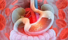
Gastrointestinal Bleeding
Gastrointestinal bleeding, is all forms of bleeding in the gastrointestinal tract, from the mouth to the rectum. Gastrointestinal bleeding is among the most common gastrointestinal disorders. Patients with GI bleeding may have different clinical presentations ranging from hematemesis or hematochezia with hemodynamic instability, to melena or rectal bleeding without hemodynamic compromise. Patients may have chronic GI bleeding with asymptomatic iron-deficiency anemia, or hemoccult-positive stool on screening for colorectal cancer.
Hematemesis is vomiting of red blood and indicates upper GI bleeding, usually from a peptic ulcer, vascular lesion, or varix. Coffee-ground emesis is vomiting of dark brown, granular material that resembles coffee grounds. It results from upper GI bleeding that has slowed or stopped, with conversion of red heamoglobin to brown hematin by gastric acid.
Hematochezia is the passage of gross blood from the rectum and usually indicates lower GI bleeding but may result from vigorous upper GI bleeding with rapid transit of blood through the intestines.
Melena is black, tarry stool and typically indicates upper GI bleeding, but bleeding from a source in the small bowel or right colon may also be the cause. About 100 to 200 mL of blood in the upper GI tract is required to cause melena, which may persist for several days after bleeding has ceased. Black stool that does not contain occult blood may result from ingestion of iron, bismuth, or various foods and should not be mistaken for melena.
Chronic occult bleeding can occur from anywhere in the GI tract and is detectable by chemical testing of a stool specimen. Acute, severe bleeding also can occur from anywhere in the GI tract. Patients may present with signs of shock. Patients with underlying ischemic heart disease may develop angina or MI because of coronary hypoperfusion.
GI bleeding may precipitate portosystemic encephalopathy or hepatorenal syndrome (kidney failure secondary to liver failure).
Causes of GI bleed are
1. Esophagitis and gastroesophageal reflux disease(GERD):- Esophagitis is an inflammation of the esophagus which damages esophagus tissue. Patients may experience problems swallowing, as well as chest pains (heartburn). It is much more common in adults than in children. GERD - known as reflux esophagitis. There is a valve which stops acids from seeping back up into the esophagus, it is called the esophageal sphincter. If it is faulty - does not flap closed and open properly, stomach contents can reflux; make their way back up into the esophagus, effectively GERD. GERD can irritate the esophagus, leading to esophagitis. Treatment is by the proton pump inhibitors and in some cases surgery is required.
2. Esophageal Varices. These are abnormally enlarged veins usually located at the lower end of the esophagus or the upper stomach. They may break open and bleed. Cirrhosis of the liver is the most common cause of esophageal varices.treatment is by banding, sclerotherapy or surgery in some cases.
3. Mallory-Weiss tear. This is a tear in the lining of the esophagus. It’s usually caused by severe vomiting. It can also happen due to things that increase pressure in abdomen, such as coughing, hiccupping, or childbirth.
4. Gastritis and ulcers:- Peptic ulcer disease is the most common cause of upper GI bleeding; over 50% of episodes are due to gastric or duodenal erosions or ulcers. GI bleeding due to ulcer disease may be brisk, subacute or chronic. In erosive disease, the mucosal defect by definition is not deep enough to result in brisk bleeding. Patients with gastric/duodenal erosions typically have modest blood loss and may present with coffee-ground emesis or melena. If gastric and/or duodenal erosive disease is diagnosed on EGD, therapy should be initiated with either proton-pump inhibitors or H2-receptor antagonists. The most common symptoms are waking at night with upper abdominal pain or upper abdominal pain that improves with eating. The pain is often described as a burning or dull ache. Other symptoms include belching, vomiting, weight loss, or poor appetite. About a third of older people have no symptoms.[1] Complications may include bleeding, perforation, and blockage of the stomach. Bleeding occurs in as many as 15% of people. Common causes include the bacteria Helicobacter pylori and non-steroidal anti-inflammatory drugs (NSAIDs). Other less common causes include tobacco smoking, stress due to serious illness, Behcet disease, Zollinger-Ellison syndrome, Crohn disease and liver cirrhosis, among others.[1][4] Older people are more sensitive to the ulcer causing effects of NSAIDs. The diagnosis is typically suspected due to the presenting symptoms with confirmation by either endoscopy or barium swallow. H. pylori can be diagnosed by testing the blood for antibodies, a urea breath test, testing the stool for signs of the bacteria, or a biopsy of the stomach. Other conditions that produce similar symptoms include stomach cancer, coronary heart disease, and inflammation of the stomach lining or gallbladder.
Diet does not play an important role in either causing or preventing ulcers. Treatment includes stopping smoking, stopping NSAIDs, stopping alcohol, and medications to decrease stomach acid. The medication used to decrease acid is usually either a proton pump inhibitor (PPI) or an H2 blocker with four weeks of treatment initially recommended.[1] Ulcers due to H. pylori are treated with a combination of medications such as amoxicillin, clarithromycin, and a PPI. Antibiotic resistance is increasing and thus treatment may not always be effective. Bleeding ulcers may be treated by endoscopy, with open surgery typically only used in cases in which it is not successful.Peptic ulcers are present in around 4% of the population.
Cause of gastric ulcers
H. pylori
A major causative factor (60% of gastric and up to 50-75% of duodenal ulcers) is chronic inflammation due to Helicobacter pylori that colonizes the antral mucosa. The immune system is unable to clear the infection, despite the appearance of antibodies. Thus, the bacterium can cause a chronic active gastritis (type B gastritis). Gastrin stimulates the production of gastric acid by parietal cells. In H. pylori colonization responses to increased gastrin, the increase in acid can contribute to the erosion of the mucosa and therefore ulcer formation.
NSAIDs
Another major cause is the use of NSAIDs. The gastric mucosa protects itself from gastric acid with a layer of mucus, the secretion of which is stimulated by certain prostaglandins. NSAIDs block the function of cyclooxygenase 1 (cox-1), which is essential for the production of these prostaglandins. COX-2 selective anti-inflammatories (such as celecoxib or the since withdrawn rofecoxib) preferentially inhibit cox-2, which is less essential in the gastric mucosa, and roughly halve the risk of NSAID-related gastric ulceration.
Stress
Stress due to serious health problems such as those requiring treatment in an intensive care unit is well described as a cause of peptic ulcers, which are termed stress ulcers
While chronic life stress was once believed to be the main cause of ulcers, this is no longer the case. It is, however, still occasionally believed to play a role. This may be by increasing the risk in those with other causes such as H. pylori or NSAID use.
Diet
Dietary factors such as spice consumption, were hypothesized to cause ulcers until late in the 20th century, but have been shown to be of relatively minor importance. Caffeine and coffee, also commonly thought to cause or exacerbate ulcers, appear to have little effect. Similarly, while studies have found that alcohol consumption increases risk when associated with H. pylori infection, it does not seem to independently increase risk. Even when coupled with H. pylori infection, the increase is modest in comparison to the primary risk factor.
Other
Although some studies have found correlations between smoking and ulcer formation, others have been more specific in exploring the risks involved and have found that smoking by itself may not be much of a risk factor unless associated with H. pylori infection.
Gastrinomas (Zollinger–Ellison syndrome), rare gastrin-secreting tumors, also cause multiple and difficult-to-heal ulcers.
Diagnosis
Diagnosis and treatment will depend on symptoms and the severity of ulcer. To diagnose a stomach ulcer, doctor will review medical history along with symptoms and any prescription or over-the-counter medications.
To rule out H. pylori infection, a blood, stool, or breath test may be done. In a breath test, one is made to drink a clear liquid and breathe into a bag, which is then sealed. If H. pylori is present, the breath sample will contain higher-than-normal levels of carbon dioxide.
Other tests and procedures used to diagnose stomach ulcers include:
=> Barium X-ray: a thick white liquid (barium) that helps the stomach and small intestine show up on X-rays
=> Endoscopy: a thin, lighted tube is inserted through the mouth and into the stomach to look for the presence of an ulcer
=> Endoscopic biopsy: a piece of stomach tissue is removed so it can be seen under microscope
Treatment
There are several ways in which ulcers can be treated, including making changes to one's lifestyle, taking medication, and/or having surgery.
Lifestyle
First treat the cause. Stop smoking, alchol and spicy diet. Include more of fruits, vegetables. Daily exercise should be done.
Medications
• Proton pump inhibitors (PPI): Proton pump inhibitors reduce acid and allow the ulcer to heal.
• Antibiotics: Antibiotics are used to treat H. pylori. There are multiple combinations of antibiotics that are taken for two weeks, along with a PPI.
Endoscopy
Some bleeding ulcers can be treated through the endoscope.
Surgery
An operation may be needed if the ulcer has created a hole in the stomach wall or if there is serious bleeding.
5. Hemorrhoids
Hemorrhoids are swollen, inflamed veins or vascular cushions (arteriovenous plexuses) in and around the anus and lower portion of the rectum. Normally they are present with cushioning from smooth vessel and connective tissue around. They become painful when swollen and inflamed.
The anal canal is the lower portion of the rectum. It is four to five centimeters in length. It begins at about the apex of the prostate or the tip of the coccyx. Here, it passes downward and backward to the anus. It is encircled throughout almost its entire extent by the internal sphincter which permits its walls to separate only during the passage of feces. The external sphincter guards its orifice, the anus. Above the outer sphincter is the levator ani muscle which supports the bowel. The pectinate line (dentate line) is a line which divides the upper two thirds and lower third of the anal canal. It lies at the inferior most level of the anal columns and indicates the junction of the superior part of the anal canal (derived from the embryonic hindgut) and the inferior part (derived from the embryonic proctodeum). Hemorrhoids are divided into two type
Internal hemorrhoids :- They are located above the pectinate line and are covered with cells that are the same as those that line the rest of the intestines.
• Grade I is small without protrusion. Painless, minor bleeding occurs from time to time after a bowel movement.
• A grade II hemorrhoid may protrude during a bowel movement but returns spontaneously to its place afterwards.
• In grade III, the hemorrhoid must be replaced manually.
• A grade IV hemorrhoid has prolapsed - it protrudes constantly and will fall out again if pushed back into the rectum. There may or may not be bleeding. Prolapsed hemorrhoids can be painful if they are strangled by the anus or if a clot develops.
External hemorrhoids:- They arise below the line and are covered with cells that resemble skin. They are very painful.
Symptoms
The most common symptom of internal hemorrhoids is bright red blood on stool.The bleeding is not severe. Internal hemorrhoids that are not prolapsed are usually not painful. Prolapsed hemorrhoids often cause pain, discomfort, and anal itching. Blood clots may form in external hemorrhoids. A blood clot in a vein is called a thrombosis. Thrombosed external hemorrhoids cause bleeding, painful swelling, or a hard lump around the anus. When the blood clot dissolves, extra skin is left behind. Excessive straining, rubbing, or cleaning around the anus may make symptoms, such as itching and irritation, worse.
Causes
Hemorrhoids are caused by repeated or constant pressure on the rectal or anal veins. The most common cause of pressure usually results from straining or prolonged sitting during a bowel movement. Other factors that increase the risk for getting hemorrhoids include constipation, diarrhea, lifting heavy objects, poor posture, prolonged sitting, pregnancy, eating a diet low in fibre, anal intercourse, and being overweight. Liver damage and some food allergies can also cause hemorrhoids.
Treatment
Simple diet and lifestyle changes often reduce the swelling of hemorrhoids and relieve hemorrhoid symptoms. Eating a high-fiber diet can make stools softer and easier to pass, reducing the pressure on hemorrhoids caused by straining. Exercise and lot of fluid prevent constipation. The medical management is
• Rubber band ligation. Special rubber band is put around the base of the hemorrhoid. The band cuts off circulation, causing the hemorrhoid to shrink.
• Sclerotherapy. Chemical solution is injected into the blood vessel to shrink the hemorrhoid.
• Infrared coagulation. Heat is used to shrink the hemorrhoid tissue.
If required surgical removal is done.
6. Anal fissures. An anal fissure (fissure-in-ano) is a small, oval shaped tear in skin that lines the opening of the anus. Fissures typically cause severe pain and bleeding with bowel movements. Fissures are quite common in the general population. The typical symptoms of an anal fissure include severe pain during, and especially after, a bowel movement, lasting from several minutes to a few hours. Patients may also notice bright red blood from the anus that can be seen on the toilet paper or on the stool. Fissures are usually caused by trauma to the inner lining of the anus. Patients with tight anal sphincter muscles (i.e., increased muscle tone) are more prone to developing anal fissures. A hard, dry bowel movement is typically responsible, but loose stools and diarrhea can also be the cause. Stool softeners, raw diet may be sufficient in most of the cases. Pain may be relieved by anaesthetic gel applications. In few cases surgery may be required.
7. Colon polyps. Colon polyps are growths on the lining of colon or rectum. Over time, some polyps can become cancerous, and removing polyps can help prevent cancer of the colon and rectum. Most people with colon polyps don’t have symptoms. It might present When colon polyps do cause symptoms, one may have bleeding from rectum, blood in stool, or feel tired because of anemia. There are different types of colon polyps with differing tendencies to become malignant and abilities to predict the development of more polyps and cancer. It is important to recognize families with members who have familial genetic conditions causing polyps because some of these conditions are associated with a very high incidence of colon cancer, and the cancer can be prevented or discovered early. Colon polyps are diagnosed by endoscopic colonoscopy, virtual colonoscopy, barium enema, and flexible sigmoidoscopy. Colon polyps are treated by endoscopic removal and occasionally by surgery. Follow-up surveillance of patients with colon polyps depends on the presence of a family history of cancer, the number of polyps that are found, the size of the polyps, and the polyps' histology, and can vary between three and ten years. Treatments to prevent colon polyps are being pursued actively.
8. Ulcerative colitis. Ulcerative colitis is a chronic disease of the large intestine, also known as the colon, in which the lining of the colon becomes inflamed and develops tiny open sores, or ulcers, that produce pus and mucous. The combination of inflammation and ulceration can cause abdominal discomfort and frequent emptying of the colon.
Ulcerative colitis is the result of an abnormal response by your body's immune system. Normally, the cells and proteins that make up the immune system protect from infection. In people with Inflammatory bowel disease , however, the immune system mistakes food, bacteria, and other materials in the intestine for foreign or invading substances. When this happens, the body sends white blood cells into the lining of the intestines, where they produce chronic inflammation and ulcerations. It may present with blood in Stools,Mucous in stools, Loose stools, Joint pains and swelling, Weakness, lethargy, shortness of breath after a walk because of anemia. The correct diagnosis is the 1st step to right treatment. All patients with blood in stools and diarrhea are not necessarily suffering from ulcerative colitis, which has to be differentiated from other diseases. COLONOSCOPY is performed to arrive at the correct diagnosis. Ulcerative colitis can occur in people of any age. However, it is more likely to develop in people between the ages of 15 and 30,older than 60, who have a family member with IBD of Jewish descent. Ulcerative colitis affects many parts of the body outside the intestinal tract.
Medication
Ulcerative colitis can be treated with a number of medications, including 5-ASA drugs such as sulfasalazine and mesalazine. Corticosteroids such as prednisone can also be used due to their immunosuppressing and short-term healing properties, but because their risks outweigh their benefits, they are not used long-term in treatment. Immunosuppressive medications such as azathioprine and biological agents such as infliximab and adalimumab are given only if people cannot achieve remission with 5-ASA and corticosteroids. This is because of their possible risk factors, including but not limited to increased risk of cancers in teenagers and adults, tuberculosis, and new or worsening heart failure (these side effects are rare).
Surgery
The gastrointestinal aspects of ulcerative colitis can generally be cured by surgical removal of the large intestine, also known as a colectomy. This procedure is necessary in the event of: exsanguinating hemorrhage, frank perforation, or documented or strongly suspected carcinoma. Surgery is also indicated for patients with severe colitis or toxic megacolon. .
9. Crohn's disease Crohn’s disease can involve any part of the gastrointestinal tract from the mouth to the anus but most commonly affects the small intestine and/or the colon. There may be areas of healthy intestine between areas of diseased intestine. Within a diseased section, Crohn’s disease can affect all layers of the intestinal wall (i.e. not just the lining). This can lead to the development of complications that are specific to this condition:
a. strictures (intestinal obstruction or narrowing of the intestinal wall)
b. abscesses (boils) and skin tags (swollen lumps or ‘flaps’ of thickened skin occurring just outside the anus)
c. fistulae (abnormal channels connecting different loops of intestine to itself or to other body organs)
d. fissures (ulcerated tears or cracks in the lining of the anal canal) malabsorption and malnutrition
SURGERY
In some cases, surgery may be required to correct the complications of Crohn’s disease or to remove diseased portions of the gastrointestinal tract (resection). Even if diseased parts of the intestines are removed, inflammation can re-appear in other areas.
Smoking increases the need for surgery and medications, can make the disease more active and may prevent the induction of remission. After surgery for Crohn’s disease, the condition may recur sooner, and often more severely in smokers than in non-smokers.
DIET & NUTRITION
Crohn’s disease in the small intestine can impair the digestion and absorption of essential nutrients from food. A well-balanced and nutritious diet is essential for anyone with Crohn’s disease in order to prevent malnutrition and maintain good health. And it is even more so for growing children and adolescents who may experience delayed growth or pubertal development in the absence of adequate nutrition.